In my Fundamentals of Neuroscience class, they are teaching us a bit of history along the way. How DID they first measure action potentials, or, even farther back (late 1800s), how DID they begin to piece apart what was happening at the cellular level in the nervous system?
We were told about Golgi's stain, which happened to stain one cell at a time, letting us see one whole neuron for the first time, and about Ramon y Cajal's work with that stain, discovering the directionality of the connection from one neuron to another.......
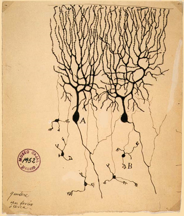
In the Understanding the Brain class I took a while back, Dr. Mason showed us a stained Purkinje cell, as one of her favorite stained cells.
When they showed us this image in Fundamentals of Neuroscience, I thought -- Hey, I bet those are Purkinje cells! And sure enough.
This image is on the Wikipedia Purkinje cell page.

"PurkinjeCell". Licensed under Public Domain via Wikimedia Commons.
Purkinje cells are in our cerebellums. Our cerebellums enable us to make smooth movements (like reaching out for a glass of water, and bringing that glass to our mouths rather than knocking it over or spilling it in our laps). Our cerebellums are constantly monitoring every small part of every movement, comparing what's actually happening to the instruction that was issued by the forebrain, and making small corrections so that the actual movement is as close as possible to the desired intent.............

"Diagram showing the brain stem which includes the medulla oblongata, the pons and the midbrain (2) CRUK 294" by Cancer Research UK - Original email from CRUK. Licensed under CC BY-SA 4.0 via Wikimedia Commons.
.


No comments:
Post a Comment